−Table of Contents
Bacterial Diseases
Mycobacteriosis (Fish TB)
Chronic, systemic bacterial infections by various Mycobacterium species are frequently diagnosed in aquarium fishes. The species most commonly found in fishes are M. marinum, M. abscessus, M. chelonae, and M. fortuitum. These species, as well as M. peregrinum and M. haemophilum, have been associated with mycobacteriosis in zebrafish (Astrofsky et al. 2000; Kent et al. 2004).
Usually mycobacteriosis is chronic with low level mortality, but in certain circumstances, infections may be very acute and result in severe epizootics in zebrafish colonies. Factors that are most responsible for the differences seen in the severity of outbreaks in fish mycobacteriosis have yet to be determined. It is not know if highly virulent strains or species cause the acute outbreaks, or if the outbreaks occur in fish that are immunocompromised by other factors, such at poor water quality, excessive crowding and handling, etc.
Mycobacterium spp. of fishes can infect humans. In most cases, the infections are confined to the extremities, and though not usually life-threatening may require aggressive antibiotic treatments to resolve (Kern et al. 1989; Shih et al. 1997; Hoyen et al. 1998). However, these infections may be lethal to immune compromised individuals (Lessing et al. 1993). Considering the zoonotic potential of Mycobacterium spp. from fishes, those handling aquarium fishes, including zebrafish, should wash their hands after coming in contact with water containing fish, and avoid exposing open lesions to aquarium water and fishes. Most cases of human mycobacteriosis associated with fishes are caused by M. marinum. This species is considered not to grow, or to grow very poorly, at 37 C. The majority of M. marinum cases are not life threatening and are confined to extremities, where body temperatures are lower. However, some strains of M. marinum are capable of growth in culture at 37 C, and we demonstrated that others not capable of growth at this temperature in media can grow at 37 C in macrophage cell lines and in mice (Kent et al. 2006).
Clinical Signs and Gross Pathology
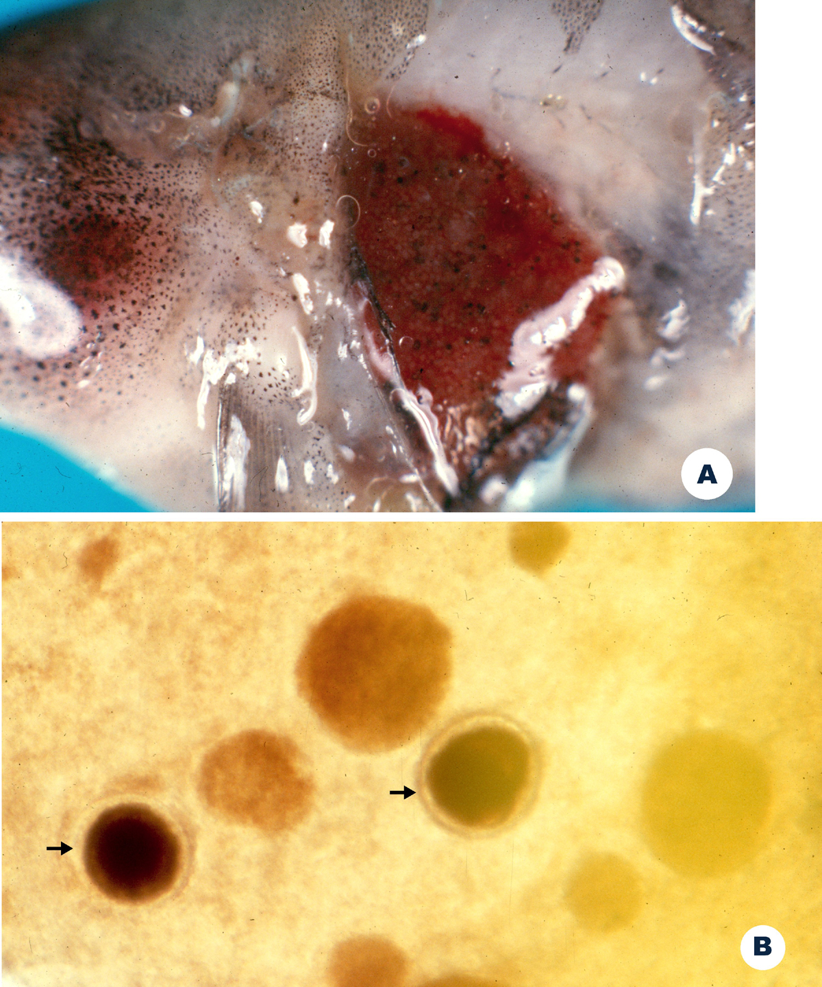
Clinical and macroscopic changes associated with Mycobacterium may be variable, dependent upon the site and extent of the infection, and on the “chronicity” of the infection. Clinical signs include lethargy, anorexia, and emaciation. Fish may exhibit ulcers, hemorrhage or hyperemia around the head (similar to Gram negative septicemia), raised scales, frayed fins, and pallor of the skin or gills. A hallmark macroscopic change is the presence of multiple, white nodules in various visceral organs. These are visible by close examination of organs with a dissecting microscope, but may be hard to find in small fish, such as zebrafish.
Astrofsky et al. (2000) described various forms of the disease as they occur in zebrafish. Infected fish showed decreased overall reproductive capabilities, as well as characteristic changes such as dropsy (generalized edema with swollen abdomens) and frank ulcers of the skin.
Microscopy
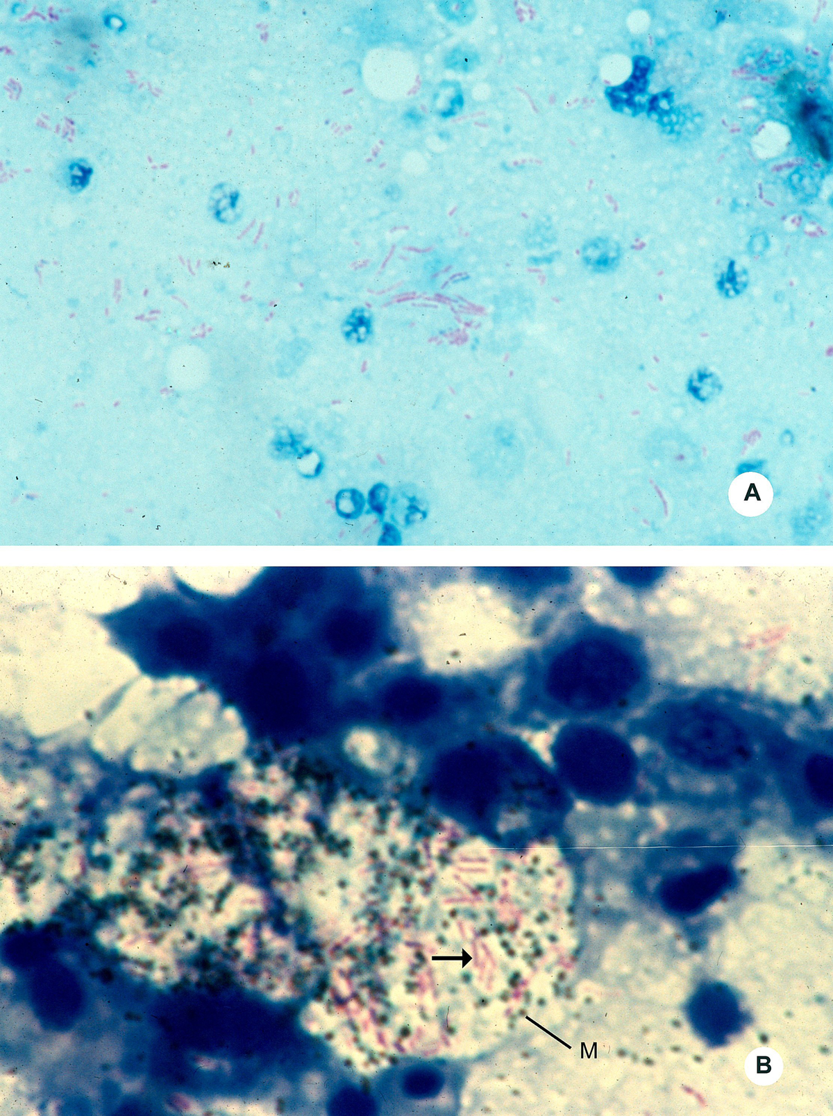
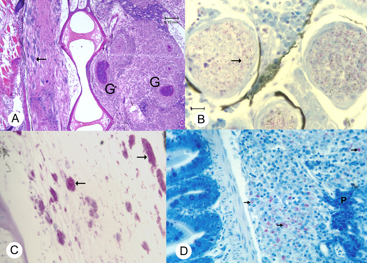
In “typical” fish mycobacteriosis (i.e., the chronic form), histological examination reveals numerous granulomas, often with necrotic centers, throughout the visceral organs. These can be seen in wet mount preparations, but can be confused with encysted parasites. Staining of these tissues with acid-fast stains will often reveal acid-fast (red) bacilli in the lesions. In more aggressive infections, associated with rapid mortality, we have seen massive accumulations of macrophages replete with bacteria throughout the viscera (severe, chronic peritonitis) and occasionally extending into the muscle. Some of these cases are very acute and exhibit few, if any, granulomas. We often visualize mycobacteria in the swim bladder wall and intestinal epithelium, indicating that these are likely sites for initiation of infections.
Diagnosis
Preliminary diagnosis is achieved by observing multiple granulomas in visceral organs in either wet mounts or histological sections. These may also be caused by fungal or parasitic infections. Confirmatory diagnosis is requires visualization of acid fast bacilli in either histological sections or in tissue imprints. Often zebrafish are decalcified during processing for histology, and we recently found that certain aggressive decalcification procedures may inhibit the acid-fast staining of Mycobacterium spp. in tissue sections (Kent et al. 2006).
Some Mycobacterium species are relatively difficult to grow in culture, and thus this procedure is infrequently employed with fish.
PCR tests for screening for the presence of the causative agent have been described (Colorni et al. 1994; Astrofsky et al. 2000). Such tests, while extremely sensitive, usually do not provide information on disease status or the severity of infection, but may be useful for screening for carrier/subclinical infections. We have also used PCR tests to obtain mycobacteria sequence directly from infected fish, which is very useful for species identification of bacteria that are difficult to grow in culture (Kent et al. 2004; Poort et al. 2006).
Control and Treatment
The infection is usually very difficult to eradicate in fish culture systems with antibiotics. However, Conroy and Conroy (1999) reported that kanamycin at 50 ppm with 4 doses 2 days apart apparently controlled the disease in guppies. Boos et al. (1995) treated firemouth cichlids and Congo tetras orally with rifampicin and tetracycline with some success. Nevertheless, depopulation of affected tanks and avoiding cross contamination has been the most effective way for controlling the infection in aquarium fishes (Astrofsky et al. 2000).
Because the disease is rather insidious, the infection is wide spread in aquarium fishes, and treatment with antibiotics is difficult, the best approach is to avoid the infection. This is accomplished by strict quarantine procedures and understanding a thorough history of the stocks introduced into research facilities. In addition, optimizing water quality and husbandry conditions will reduce the likelihood of epizootics occurring when a few carry fish are present in the population.
Gliding Bacteria
Several species of gliding bacteria (i.e., Flavobacterium, Flexibacter or Cytophaga spp.) caused surface infections in freshwater and marine fishes. One of these diseases, bacterial gill disease (BGD), is an infection caused by a variety of these bacteria, precipitated by crowding and poor water quality. Therefore, BGD is also called “environmental gill disease”. Two gliding bacteria, Flavobacterium columnare and F. branchiophilum, are species specifically incriminated with the disease in freshwater. Other bacteria may cause or be secondarily involved with the lesions. Key predisposing factors are high organic loads and possibly high ammonia in the water. Therefore, the condition is often found in high production trout farms. With zebrafish, we have seen BGD following shipment in which the fish were in transit for a long time.
Clinical Signs and Gross Pathology
Typical of most gill diseases, fish exhibit labored breathing and may accumulate at the surface with BGD. Fin and tail infections (known as fin and tail rot), is characterized by erosion of the fins. Infected skin usually appears white due to loss of the overlying epidermis and exposure of the dermis.
Microscopy
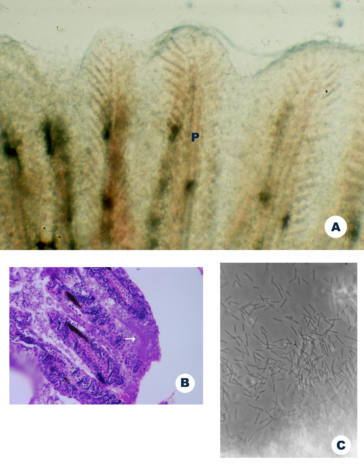
Wet mount examinations of infected gills reveal fused secondary lamellae due to epithelial hyperplasia. Masses of filamentous bacteria are observed on the surface of the gills. Likewise, masses of bacteria are seen in wet mount preparations of infected skin or fins. Phase contrast microscopy is useful for visualization of the bacteria. Histological sections reveal severe epithelial hyperplasia of the gills and occasionally necrosis. Careful examination usually reveals mats of bacteria on the gill surface.
Diagnosis
Gliding bacteria infections are usually diagnosed by observing masses of bacteria from the infected tissue. With BGD, the gill surface is associated with excessive mucus production and hyperplasia of the gill epithelium. All of these changes can be visualized in wet mount preparations of the infected tissues. Histology is useful to demonstrate pathological changes of the gills and the extent of invasion by the bacteria in the skin.
Control and Treatment
The best way to avoid BGD and fin and tail rot is to avoid overcrowding and maintain proper water quality. When shipping, assure that fish are not feed for a few days before transport, maintain lower densities in shipping bags, and select the fastest route of travel.
The most common chemical treatment for BGD in salmonids has been chloramine T at about 6-15 ppm as a 1h bath (Bowker and Erdahl 1998). Repeated treatments may be required. Lumsden et al. (1998) compared this drug with hydrogen peroxide for treating experimental infections in trout, and ultimately recommended 100 mg/L hydrogen peroxide (1 h baths) as an effective treatment as well. However, we have not tested hydrogen peroxide on zebrafish.
Bacterial aerocystitis (swim bladder bacterial infections)
M. Kent1; J. Matthews1, E. Laver2; J. M. Schech2
1. Zebrafish International Resource Center
2. National Institutes of Health, NICHD, Bethesda, MA 20892
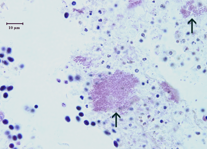
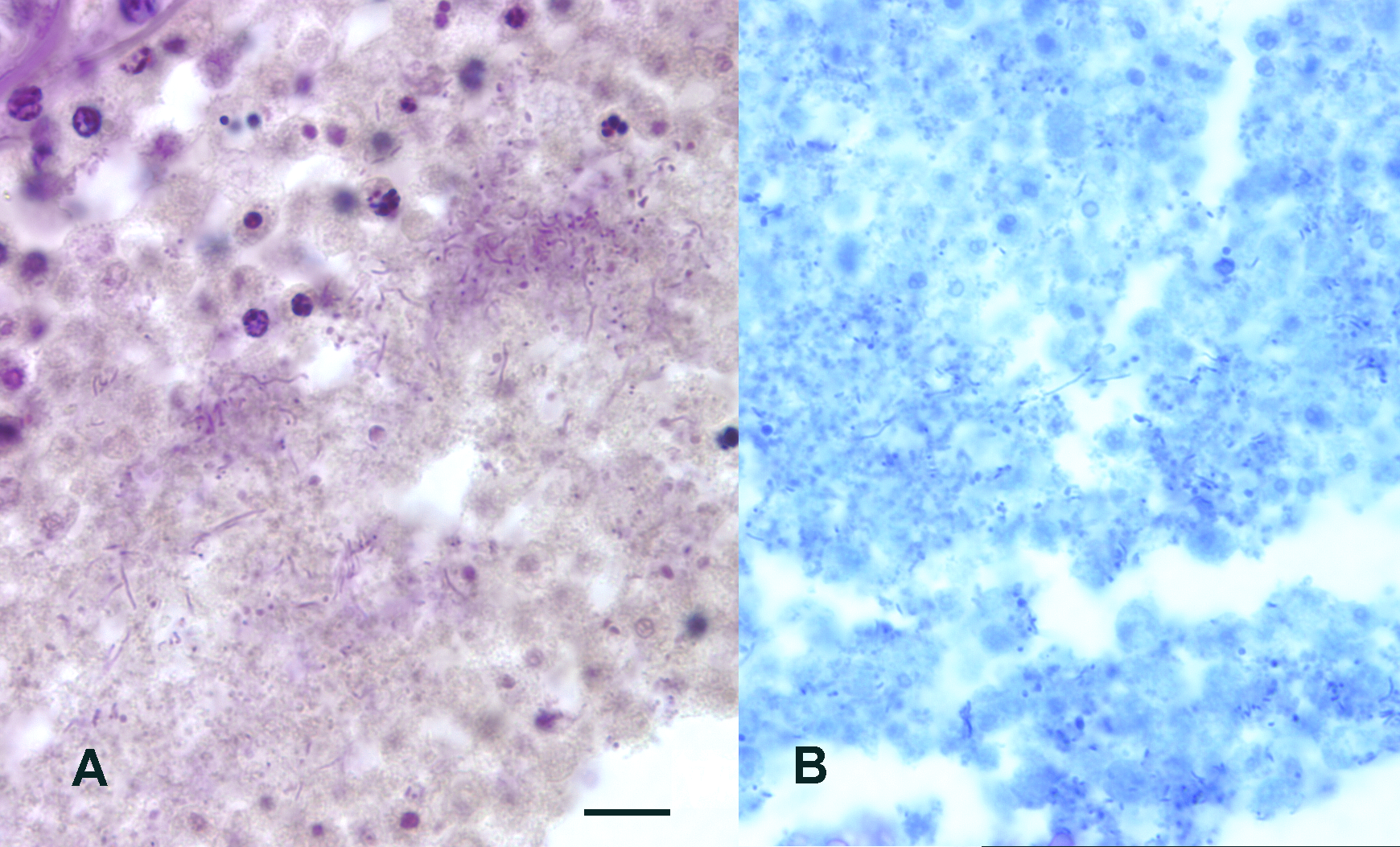
Bacterial and fungal infections of the swim bladder are occasionally reported in various fish species (Aho et al. 1988; Blaylock et al. 2001; Bowater et al. 2003: Bruno 1989; Lehmann et al. 1999; Miyazakai et al. 1984;Wada et al 1993). We have observed severe, chronic bacterial infections of the swim bladder (aerocystitis) at three zebrafish facilities, and one of these suffered very high mortality associated with the condition. Cultures of the kidney (the organ routinely cultured for systemic infections in fish) were inconsistent. At one facility, most fish were negative for routine bacterial culture of the kidney, while a few others showed mixed infections by Aeromonas hydrophila, Aeromonas sp., and Pseudomonas sp. Similar bacteria were isolated from the visceral cavity. Dr. Ana Baya, Maryland Department of Agriculture isolated Vibrio cholerae non 01 from fish from the same outbreak. Examination of histological sections with special stains reveal a variety of bacteria types. Where as typical bacilli are seen in most cases, large numbers of long, filamentous bacteria were observed associated with the lesions at one facility. Miyazakai et al. (1984) isolated Pseudomonas fluorescens from inflamed swim bladders of tilapia.
During capture a hypodermic needle is often inserted into the swim bladder of physoclistous fishes (fish with closed swim bladders) from deep water for degassing purposes (Blaylock et al. 2001; http://www.soest.hawaii.edu/SEAGRANT/opakapaka/opakapaka.html). The underlying cause of this condition in zebrafish has not been determined. Zebrafish have an open swim bladder that connects to the gastro-intestinal tract (physostomous), which would allow for gut bacteria to colonize the swim bladder of a susceptible (e.g., immunocompromised) host. At least at one facility the affected fish were not subjected to known stressors such as handling (e.g., squeeze spawning) before the onset of the disease. Interestingly, many fish from the most severe outbreak exhibited exceptionally severe microsporidiosis of the muscle. Given that microsporidia are well-recognized causes of opportunistic infections in immunosuppressed hosts, that mixed bacteria were cultured, and that no iatrogenic causes for the swim bladder infections was identified, the possibility exists that the fish become susceptible to aerocystitis after becoming immunosuppressed by unknown agents or poor water quality.
Clinical signs and gross pathology
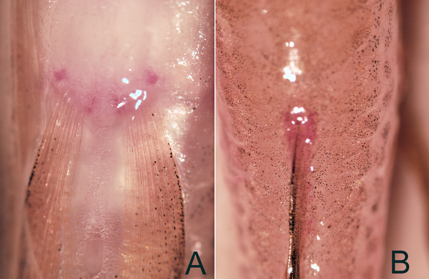
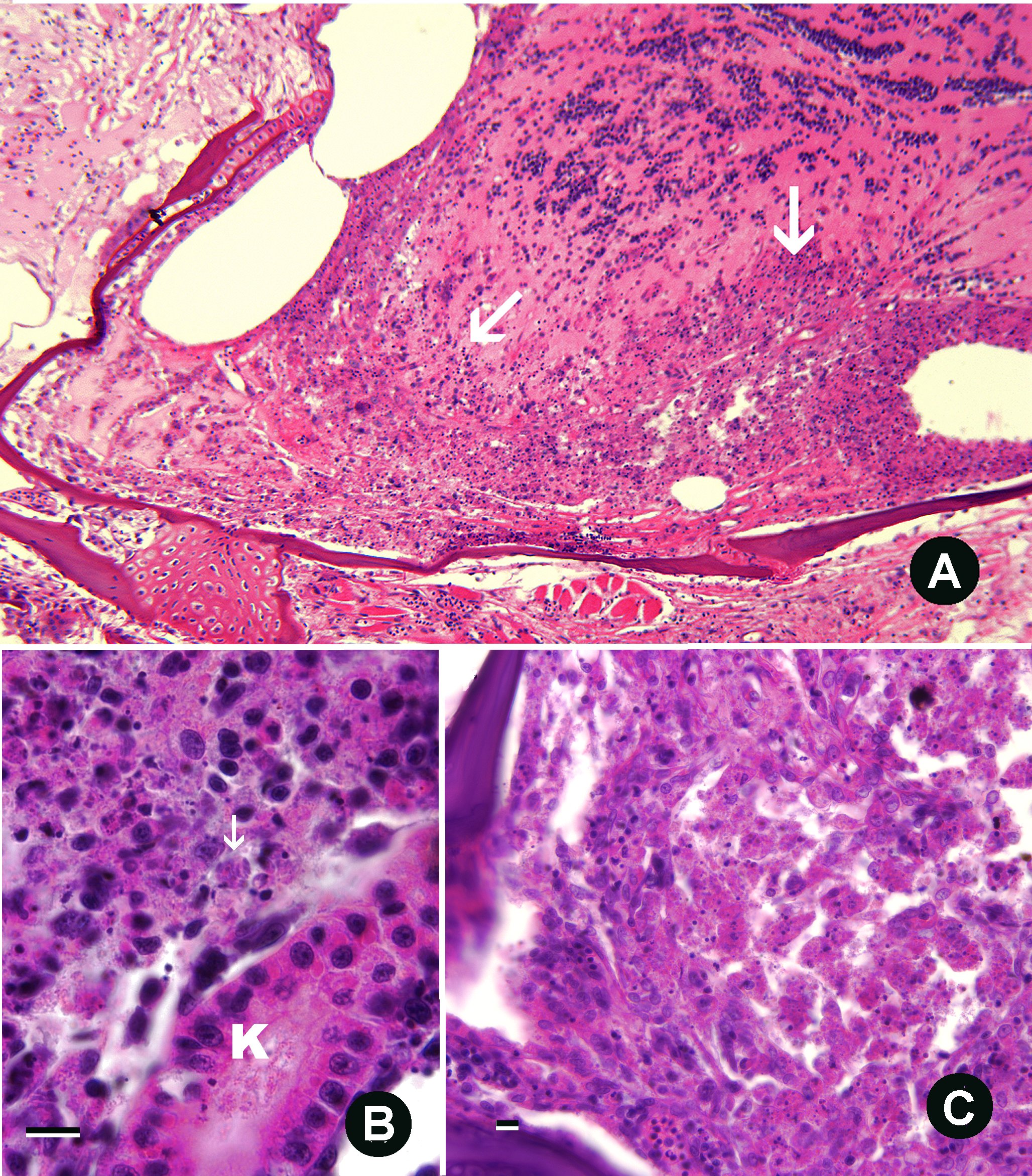
Affected fish may be lethargic and accumulate near the bottom of the tank, which is consistent with impairment of the swim bladder. Fish often exhibit erythema at the base of the fins (see below). It should be noted that hemorrhaging and erythema at of the skin and base of the fins is also characteristic of Gram negative septicemia and viral disease, and is thus not pathognomonic for this condition.
Microscopy
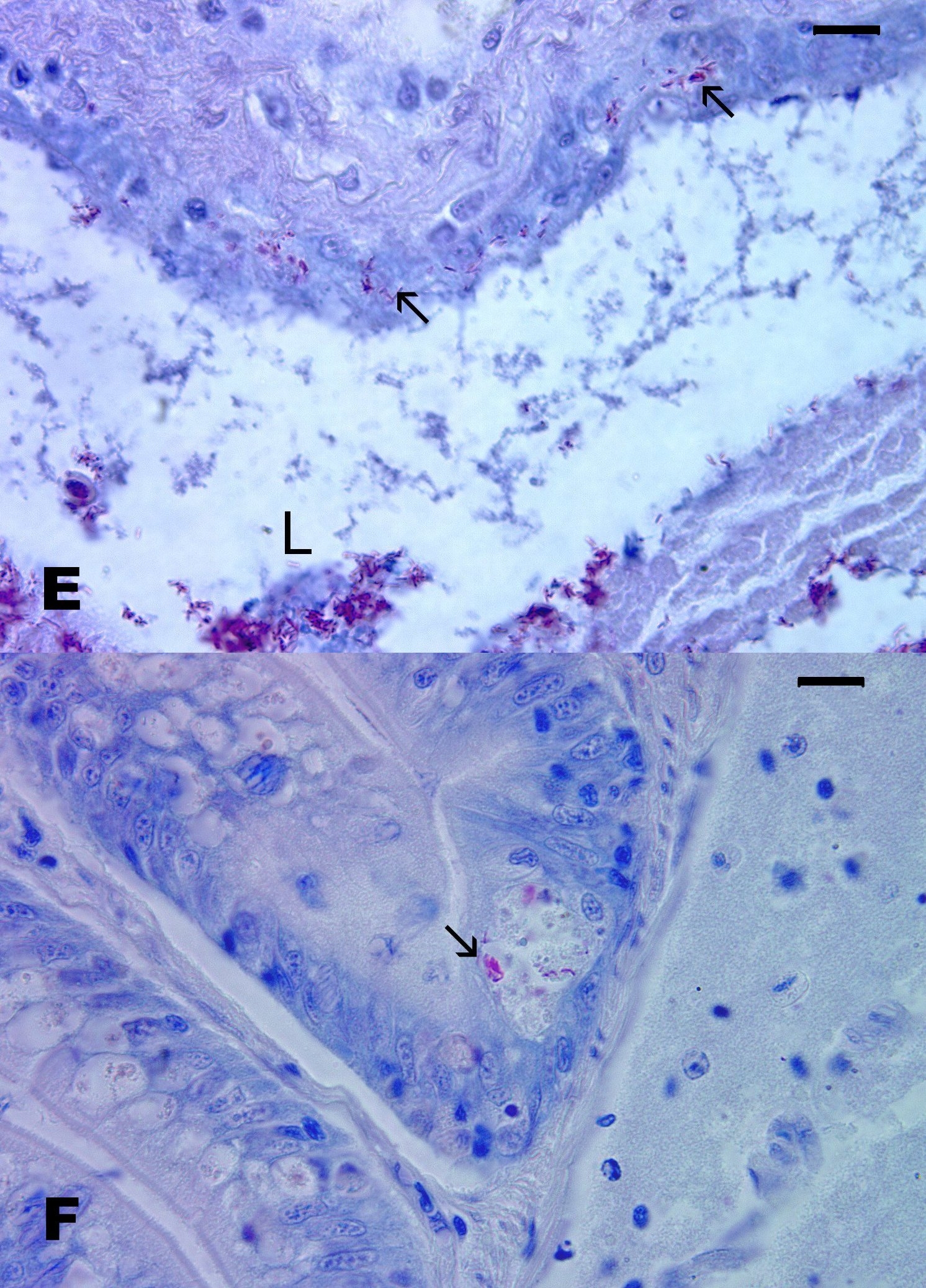
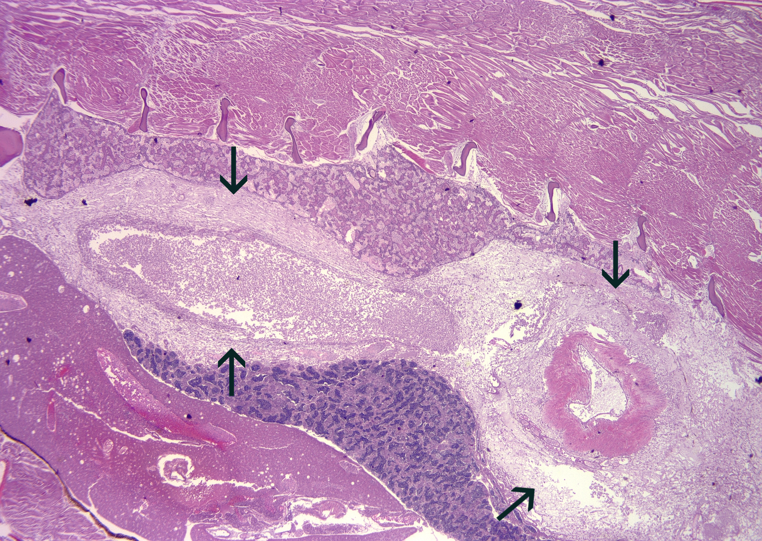
Histological evaluation revealed massive destruction and severe, chronic inflammation of the swim bladder (see below). The lesion often extended throughout the visceral cavity and dorsally through the kidney into the somatic muscle. High magnification of tissue sections revealed prominent necrosis and Gram stains of sections often showed large colonies of bacteria (see below). Gram stains may not always demonstrate bacteria in cases where filamentous bacteria are involved. These bacteria, also referred to as gliding bacteria, belong to the genera Flexibacter, Flavobacterium, or Cytophaga. We stained sections with an acid-fast stain that incorporated methylene blue as a counter stain, and massive numbers of blue-staining (acid-fast negative) bacteria where visualized (see figure). However, bacteria were inconsistently observed using various methods of the Gram stain. The Steiner sliver stain is also usual for demonstrating these types of bacteria in sections. Kent et al. (1987) found that Flavobacterium psychrophilum could not be readily seen in sections stained with hematoxylin or eosin or Gram, but were visible with Giemsa stain.
Diagnosis
Macroscopic changes could be confused with traditional Gram negative septicemia. Diagnosis, therefore, is based on observing the characteristic lesions in the swim bladder by histology. Various special stains (including Gram, Steiner or methylene blue) should be considered for demonstrating bacteria. Bacterial culture is unreliable as a specific bacterial species causing the condition has not been identified.
Control
We have yet to identify the underlying cause of this condition. It is likely that the bacteria are opportunists as the infection is associated with several different bacterial species. Until the causes are of aerocystitis is unidentified, we recommend keeping affected fish isolated from other populations and assuring that water quality is optimum.
Edwardsiella ictaluri
This Gram negative is a serious disease of channel catfish in aquaculture (Plumb 1999). Zebrafish are very susceptible, and hence have been used as a vaccine model (L. Petrie-Hanson et al. 2007). Zebrafish are generally quite resistant to opportunistic Gram negative bacteria, such as Aeromonas and Pseudomonas species, but E. ictaluri doesn’t fall into this category. It is a primary pathogen, and unlike aeromonads, etc., merely placing the bacterium in water with zebrafish results in disease (L. Petrie-Hanson et al. 2007). Natural transmission occurs primarily through shedding of bacteria from carriers or diseased fish, or by cannibalism of dead fish. In other words, it is thought that the main source of infection is infected fish, rather than the environment. However, in catfish the bacterium is capable of persisting in the environment without fish once it has been introduced (Plumb 1999). Working with colleagues, we recently diagnosed outbreaks of this infection at three separate facilities, two in fish held in quarantine and one in a main facility that used the “eggs only” policy. Although considered primarily a disease of channel catfish, the infection has been reported in other freshwater fishes (Plumb 1999). It has been observed in aquarium fishes used in research at universities in the past, including in the green knife (Kent and Lyons 1982) and Danio devario (Waltman et al. 1985).
Clinical Signs and Gross Pathology
Typical of Gram negative septicemias in fish, infected fish exhibited high mortality and diffuse erythema (reddening) of the skin. Whereas high mortalities often occur following exposure, studies with catfish showed that some fish may remain carriers for many months (Plumb 1999).
Microscopy
Infected fish exhibit severe necrosis in visceral organs, particularly in the spleen and kidney. Fish also show a severe, locally extensive necrosis and inflammation of the forebrain, olfactory nerves, and the nares. The nares have been shown to be an important route of infection in catfish (Plumb 1999). High magnification examination of zebrafish sections reveals numerous bacterial rods in phagocytes in these lesions, as well as the kidney and spleen.
Diagnosis
Presumptive diagnosis is obtained with H&E stained sections, and is based on observing necrotic changes in the forebrain, nerves and nares, with numerous bacilli in phagocytes. Confirmatory diagnosis is obtained by culture and biochemical identification of bacteria (Hawke et al. 1981; Waltman et al. 1986). The bacterium is more fastidious and grows slower in culture than many other Gram negatives found in fish. Co-infections with Aeromonas hydrophila have been seen in catfish (Nusbaum and Morrison 2002), and we have seen this in zebrafish. This may make diagnosis more difficult as A. hydrophilia grows much more rapidly in culture than E. ictaluri. Edwardisella ictaluri grows well on blood agars and brain-heart-infusion agar. We held cultures from zebrafish at 25-30 C, but 5-7 days were required to visualize prominent colonies. So far the strains that we have isolated from zebrafish are not motile, which is distinct from most of the catfish strains. Further confirmatory diagnosis can be obtained by amplification of the small subunit rDNA (AF310622) and sequencing of the PCR product.
Control and Treatment
The best approach is to avoid introducing infected fish, using quarantine, and “eggs only” methods. Fortunately, the infection does not appear to be widespread in zebrafish, and two of the outbreaks were identified and stopped in quarantine before fish were introduced to a main facility. The infection is common in channel catfish and has been reported in pet fish species. Therefore, particular caution should be used when researchers obtain fish from pet fish dealers. As the bacterium is very pathogenic and not widespread in zebrafish, we recommend that infected populations be euthanized and all equipment be disinfected. Antibiotics used to control the infection in catfish include oral administration of oxytetracylcine or ormetoprim-sulfadimethoxine. Other drugs that have been tested in vitro include kanamycinm, streptomycin, and oxolinic acid (Noga 2010)
