Table of Contents
Starvation Syndrome
Severely emaciated fish show a common collection of pathologic changes in the muscle, liver, intestine, and ovary. The cause is one of many underlying known or unknown chronic diseases resulting in this overall presentation of cachexia, a complicated metabolic syndrome related to underlying illness and characterized by muscle mass loss and is often linked to anorexia.
Clinical Signs and Gross Pathology
Affected fish are very emaciated, and usually are notably lethargic.
Microscopy.
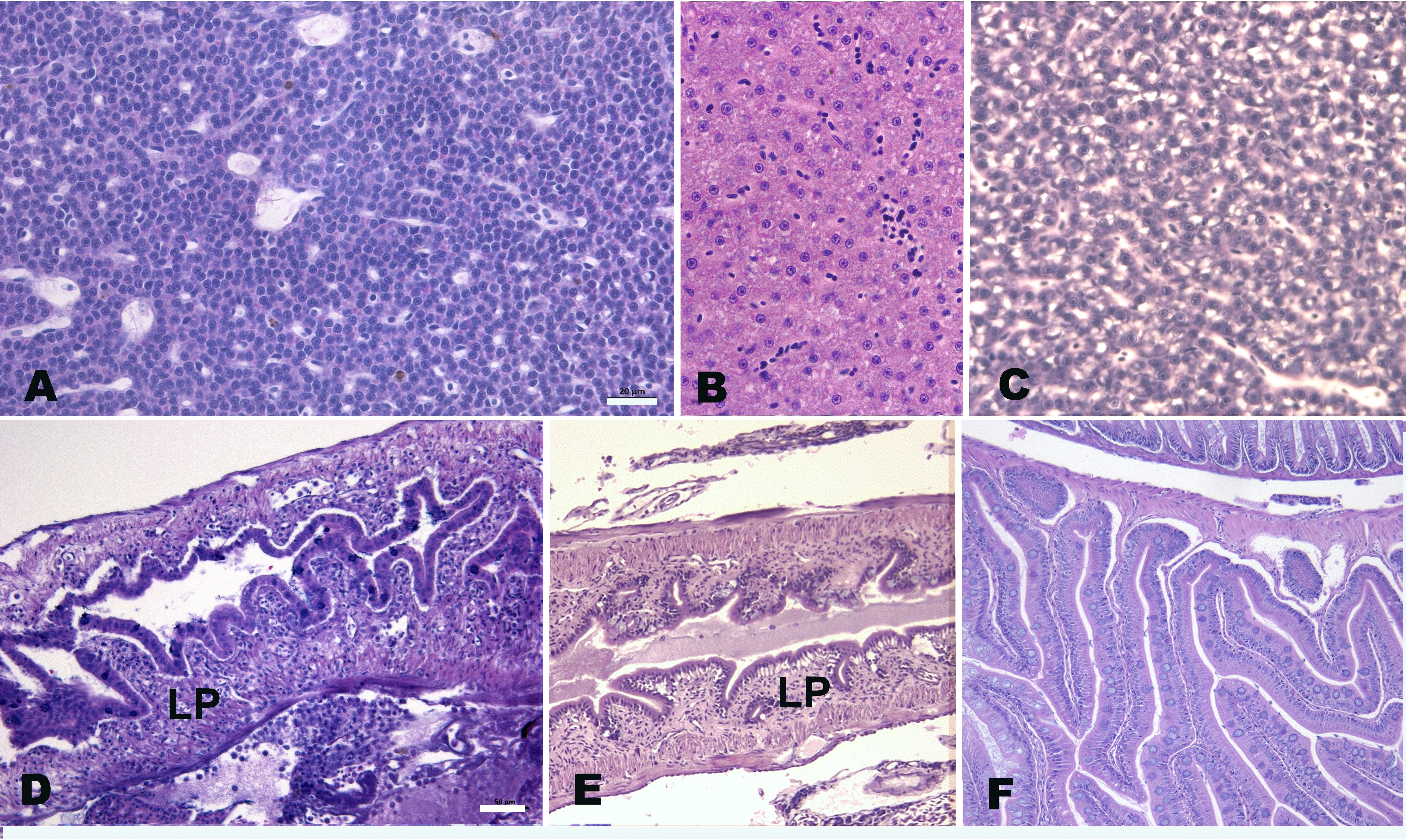
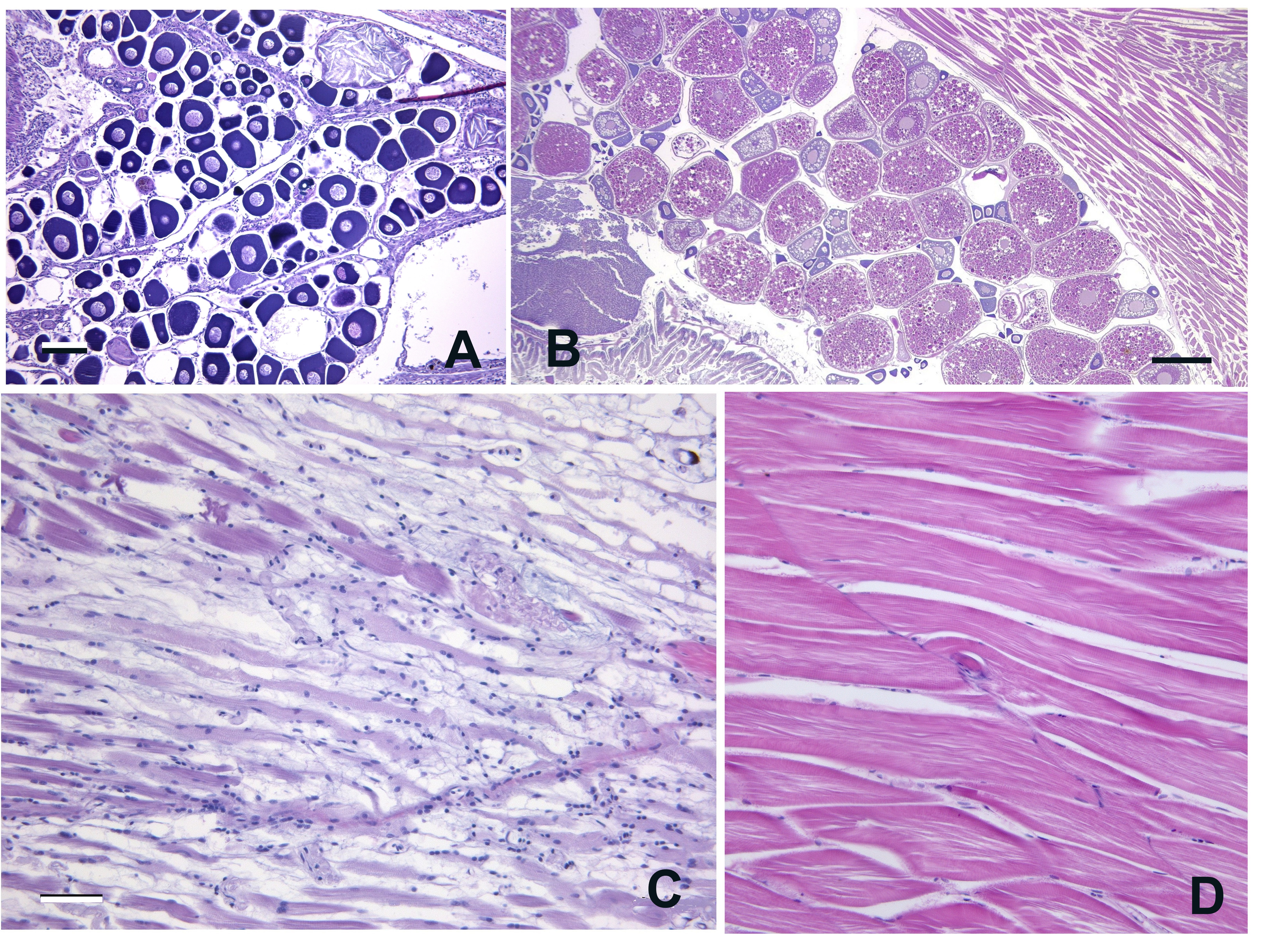
The entire intestine shows atrophy and reduction in height of the plates, consistent with villar atrophy seen in mammals, and chronic inflammation of the lamina propria. The muscle often shows severe, diffuse myodegeneration. The liver exhibits severe reduction in the cytoplasm to nucleus ratio of essentially all hepatocytes, to the extent that at first appearance it is may be difficult to recognize as liver. With females, the ovary is atrophied and no large, vitellogenic eggs are present – i.e., only small, earlier stage oocytes are seen. Diagnosis. Severe emaciation (thinness) is a useful preliminary diagnosis. Confirmation is made by observation of the pathologic changes in histological sections.
Diagnosis.
Severe emaciation (thinness) is a useful preliminary diagnosis. Confirmation is made by observation of the pathologic changes in histological sections.
Control and Treatment
Starvation syndrome is the result of many chronic diseases, as well as some unknown causes. As seen with cachexia in humans and other higher vertebrates, the presentation is the end result of long standing diseases such as chronic infections or cancer. We frequently have seen this effect some fish within a population afflicted with intestinal cancer, Pseudocapillaria tomentosa infections (Paquette et al. 2013; Gaulke et al. 2019), other neoplastic diseases, severe mycobacteriosis, or chronic gill disease. Regarding the latter, it is unknown if starvation syndrome is the result or the predisposing cause of gill disease when lethargic fish are ataxic at the bottom of the tank.
The cause of SS is often unknown as the fish present with no other obvious pathologic conditions. Similar changes are seen in the intestine in severely immune compromised humans with AIDS or Common Immune Deficiency Syndrome, suggesting that an idiopathic immunosuppression agent should be considered. Anorexia is probably an important component of SS, but we have seen that simple fasting, even for a month, does not induce the disease.
Figure 1. Liver and intestine of fish with Starvation Syndrome (SS) compared to normal. H&E. A. Liver of fish with SS. Note greatly reduced cytoplasm to nucleus ratio in all hepatocytes. Bar = 20 µm. B. Liver from normal male, with more prominent cytoplasm in hepatocytes. C. Liver from normal female, with typically more basophilic cytoplasm compared to males. D, E. Intestines of two fish with SS. Note reduced height of intestinal plates and prominent, diffuse, chronic inflammation of underlying lamina propria (LP). Bar = 50 µm. F. Intestine from normal fish with prominent intestinal plates.

Figure 2. Ovary and muscle of fish with Starvation Syndrome and normal fish. H&E. A. Atrophy of the ovary in a fish with SS. Note only immature oocytes and no vitellogenic eggs. Bar = 100 µm. B. Normal ovary (lower magnification), replete with large, eosinophilic, vitellogenic eggs. C. Diffuse myodegeneration and myositis in fish with SS. Bar = 50 µm. D. Normal muscle.
